
DEFINITION OF NEURAL TUBE DEFECT
These are broad-spectrum congenital anomalies that occur as a result of a closure error in the brain,spinal cord and spine in the first weeks of embryonal life. Closure of the neural tube occurs irregularly in both head and tail directions in 5 different regions at the same time.
Classification of neural tube defects:
- Anencephaly
- Spina Bifida
- Encephalocele
- Inencephaly
- Craniorasis
SPINA BIFIDA DEFINITION
Spina bifida, which is a congenital problem, is caused by the closure problem of the neural tube and occurs in the embryological period . It is the second most common cause of childhood disability after cerebral palsy . Hippocrates defines Spina Bifida as : “A disease that descends from the head into the spinal cord with veins and attacks the sacral bone .”

Picture 1. Meningomyelocele patient figures excavated in anthropological excavations
The causes of Spina Bifida include the lack of active substances during the closure of the spine in the 4th week of pregnancy, such as folic acid, or the drugs used. Insulin-dependent diabetes , epilepsy, genetic variations and obesity increase the risk of spina bifida in the mother.
The classification of spina bifida was made according to whether the nerve structures emerged from the disorder in the spine and whether they were covered with normal skin structure. Spina Bifida can be classified as SB Okulta, SB Menifesta, SB Aperta and SB Sistika.
- Spina Bifida Okulta: Althoughthere is a problem in the development of the vertebra, the spinal membranes or the spinal cord do not protrude. The lesion is covered with skin. There may be hairiness, dark spots, dimples or slight swelling on the skin . They usually do not show any symptoms .
- Spina Bifida Menifesta:There are findings such as open or closed nerve structures and hemangioma, hairiness or cavity.
- Spina Bifida Aperta :The area is usually reddish , translucent, and covered with a membrane that leaks cerebrospinal fluid and has a skin that surrounds the membrane. This table is also called myelosis.
- Spina Bifida Sistica: Spina Bifida Sistica is divided into two as meningocele and meningomyocele. They constitute 80% of SS cases.
A) Meningocele:Spinal fluid and membranes have overflowed without opening in the spine . This opening does not include nerve structures, and because of this feature, it is separated from myelosis and meningiocele, and may or may not be skin on the lesion.

Picture 2

Picture 3
B)Meningomyocele(MMS) : It is the most serious form of Spina Bifida. It affects the central and peripheral nervous systems. It can be explained as the formation of a space in the spine as a result of the junction error in the midline of the caudal neural tube and the overflow of the membranes and nerve structures out of the spine through this space. Neurological, orthopedic and urological problems can be seen. Hydrocephalus is one of the most common neurological complications. The higher the lesion level, the higher the risk of hydrocephalus. Urological problems can be explained as neurogenic bladder, escape from the bladder to the ureter, recurrent pyelonephritis and kidney stones .
Etiology
Although the etiology is not fully known, we can list the risk items as follows:
1.Genetic Factors
2.Familial Story
3.Folic acid
4.Diabetic Mother
5. Medicines
6. Socio-economic factors
7.Hyperthermia During Pregnancy
8.Maternal Age
9.Maternal Profession
10.Obesity
CLINIC FINDINGS
Clinical findings in Spina Bifida vary according to the location , size , and affected brain structures of the lesion.
Motor Loss : It appears as paraplegia or motor loss at different levels. Sometimes motor losses can be seen in the upper extremity. Motor losses may vary depending on the lesion level. Neuromuscular involvement in meningomyelocele can occur in three different ways. While flask paralysis, sensory and reflex loss are observed below the level in lesions resembling complete cord incision, voluntary movement or sensation may be preserved in incomplete lesions. While there is function in caudal segments in jumping lesions, there may be some non-functional segments in between.
Sensory Findings : Sensory level may not be compatible with motor level. While superficial sensory loss leads to skin problems, deep sensory loss can lead to balance and movement problems.
Osteoporosis and Fractures: In Spina Bifida, inadequacies occurs in its normal ambulation due to sensory and motor disorders in the lower extremity, and this causes osteoporosis and pathological fractures.
Hydrocephalus: Hydrocephalus is the pathological expansion of the spaces in the skull, usually the cerebral ventricles, as a result of the deterioration of the balance between the secretion and absorption of cerebrospinal fluid (CSF). Approximately 80%-90% of cases with newborn spina bifida have hydrocephalus,mostly congenital, but may also develop in the first week of life. Hydrocephalus treatment is performed by inserting a shunt in the first period of life. Generally, complications occur in the first years of life, but can also occur in any part of life. It is important to make the necessary symptoms when neurological symptoms ocur.
Orthopedic Problems: Congenital or acquired orthopedic problems can be seen. Acquired developmental deformities are associated with the level of the lesion and may occur due to muscle strength imbalance, paresis/plegia and decreased sensation in the lower extremities. Orthopedic problems may also occur due to iatrogenic (a disease that develops in the treatment of a disease due to this treatment) such as damage as a result of tetra cord surgery. The following problems can be seen mostly related to spine, hip, knee, foot and ankle structures.
1.Upper Extremity Tissue Injuries
2.Motor Problems
3.Cognitive Problems
4.Neurogenic Bladder
5.Neurogenic Intestine
6.Sexual Problems
7.Tetra Cord Syndrome
8.Vision Disorders
9.Chiari II Malformation
10.Obesity
11.Latex Allergy
PHYSIOTHERAPY EVALUATION
By evaluating the child with Bifida spina, it is aimed to determine the situation of the child at the time of evaluation, to plan the appropriate treatment program, to take precautions against secondary problems that may develop and to follow the changes that suggest progressive neurological dysfunction. Evaluation parameters can be itemized as follows.
- Patient History
- Inspection
- Posture Evaluation
- Anthropometric Measurements
- Attention, Cognition, Comprehension
- Evaluation of Cranial Nerves
- Sense Assessment
- Evaluation of Normal Joint Range of Motion
- Evaluation of Muscle Strength
- Evaluation of Neurogenic Intestine and Bladder
- Determination of Motor Lesion Level
- Tonus Assessment
- Evaluation in Terms of Orthosis and Auxiliary Devices
- Evaluation of Daily Living Activities
In spina bifida, second motor neuron findings such as muscle weakness, hypoesthesia/ anesthesia, decreased or inability to obtain deep tendon reflexes are observed. These may be accompanied by vertebral disorders such as scoliosis and kyphosis; hip dislocation, contactures in the hip and knee, ankle and foot deformities .

Picture 4
In the examination, whether there is an open, operated or skin-covered sac in the back area, increased hair growth and color changes are evaluated. Spontaneous movements in the lower extremity are observed by monitoring the babies for a while. In palpation, spinous protrusions are followed, deficiencies or abnormal formations are detected. Vertebral deformities and lower extremity contractures are disorders that negatively affect functional mobility.
TREATMENT METHODS IN PATIENTS WITH SPINA BIFIDA
SURGICAL TREATMENT
Situations requiring surgical intervention in spina bifida:
- To repair and close the lesion,
- Treatment of hydrocephalus,
- Orthopedic problems: It generally includes curvatures in the back, hip dislocation, foot-ankle deformities and also fractures.
- Bladder and bowel problems.
Pediatric, orthopedic and plastic surgery teams may be needed.
PHYSICAL THERAPY AND REHABILITATION
The aim of physiotherapy and rehabilitation in Spina Bifida is to minimize inadequacies by maximizing functions, to ensure their participation in society by making necessary changes around them, to ensure independence, to restore muscle tone to normal, to ensure that the correct posture and movement is learned from the early period, to prevent muscle and joint stiffness that may develop, and to ensure that normal posture and movement patterns are acquired.The physiotherapy and rehabilitation program to be applied to the child with spina bifida is planned according to the age of the child and the motor and sensory deficits that occur according to the spina cord deformity.
In the Newborn Period ; Treatment should be started on the day the child is born. Abnormal postures and deformities can be seen, and feformities can be congenital. For this reason, plastering and dressing are performed first. Passive movements and stretching exercises can be performed to improve joint opening and muscle length. Normal joint movements are taught to the family to prevent deformities.


Picture 5-6
Bobath Treatment; Supports normal movement development by shaping the treatment with functional activity.For babies, we can call Bobath a game concept. Toys can be used during treatment.

Picture7

Picture 8
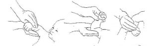
Vojta Treatment; It helps to reduce sensory disorders and muscle weakness, provides contraction in the bladder, and is effective in reducing remaining urine and preventing joint contractures.In the Vojta technique, 7 postural reflexes are defined: Vojta reflex ,Traction reflex, Peiper reflex ,Vertical collis, Horizontal collis, Landua reflex, Axillary vine response. Reflex crawling can occur by giving resistance to touch, pressure application, stretching and agonist muscles at trigger points in 3 positions (beginning, middle, end). For example, to facilitate reflex rotation and crawling, there are 9 main stimulation points such as humeral inner epicondyle, femur inner condyle, calcaneus and auxiliary stimulation points such as SIAS, acromion, scapula lower end, gluteal region.

Picture 9

Picture 10
In Preschool Treatment ; Joint range of motion is maintained and circulation is increased with passive movements. After the child starts active joint movements, he/she provides mobilization by using his/her extremities in daily life. After these movements start, necessary strengthening exercises should be given considering the weaknesses in muscle strength. By bringing the child to prone, supine and sitting positions, normal development is supported and muscle length is provided. Ensuring the free use of hands by supporting the child in the sitting position is an important point in high-level lesions. The child should be directed to the necessary assistive devices by making mobility evaluations in bed, at home and in the community. These can be items and tools such as orthoses, wheelchairs, modified bicycles. Children with heavier disabilities sit constantly because they cannot stand. In this case, the risk of developing postural instability is high. Postural symmetry should be provided to prevent this.

Picture 11-12

Picture 13
Losses and weaknesses in muscle strength, sensory disturbances and range of motion should be checked during school age. Stretching and strengthening exercises should continue. In the daily life activities of the patient, the loads on the joints during walking and transfer should be examined. Tissues should be examined and if there is pressure on the tissue, pressure reducing methods should be developed. When the child reaches school age, it is very important for the physiotherapist to help the family and educators and to arrange the home and school environment. If the child has a shunt, the entire team working with the child should be informed about this issue and should pay attention to symptoms such as headache, vomiting, decreased performance, and vision problems. In order for the child to use his/her hands more comfortably, the physiotherapist or occupational therapist should teach him/her basic wheelchair skills. During this period, he/she can be directed to sports activities such as swimming, hand cycling or wheelchair basketball. These activities increase the strengthening, increase the blood circulation and prevent the formation of pressure wounds by providing body weight control. In daily life, it is very important for the child to be in a proper posture and to transfer equal weight during the time he/she spends in a wheelchair.
Stretching exercises prevent shortness and joint contractures in hip flexors, hamstrings and gastrocnemius and soleus muscles. For the stretching of the hip flexors, the child should stand or lie face down for at least one hour a day, sit in a long sitting position with orthosis for hamstrings, and take the plantigrade position in sitting or standing position for the gastrocnemius and soleus muscles.

Picture 14
 Picture 15

Picture 16

Picture 17

Picture 18  Picture 19
 Picture 20

Picture 21
Mobility assessment should be made especially in terms of long distances inAdult Treatment. Necessary equipment should be evaluated and implemented by making necessary changes to make mobility independent and safe. The treatment program should be renewed according to changes in range of motion, muscle strength, endurance,motor skills. In order to prevent obesity and pressure ulcers, it is important that individuals maintain their form and actively continue sports clubs.
Orthoses
Duties of orthoses;
1.Supporting the body against gravity
2.Protecting the joints from pathomechanical damage
3.Increasing the effectiveness of walking
4.Prevent or reduce the development of contractures in the joints
5.To protect the bone and joint ligament after surgery
6.To provide vertical position instead of wheelchair.
Orthoses that help the child transfer weight by supporting him/her in an upright position;
1.Allow the child to perceive the vertical position
2.Increased visual perception
3.Increased communication with the environment
4.Functional use of the upper extremity
5.The development of head control
6.Improvement of body control
7.To the development of equilibrium reactions
8.Regulating the tone
9.Prevention of contractures
10.Reduction of pressure sores
11.Decrease in obesity
12.It helps to support cardiovascular, pulmonary, urinary and gastrointestinal system functions.
The plantar flexor muscle group is weak in patients with lower lumbar involvement. It is recommended to use CAFO or AFO in patients with lower lumbar involvement.

Picture 22: KAFO

Picture 23 :AFO
Patients with middle lumbar involvement often have weakness in the muscle groups below the knee. Hip extensor and abductor muscles are weak. The use of HCAFO, CAFO and condylar-supported AFO is recommended in patients with moderate lumbar involvement.

Picture 24 : HKAFO
Knee extensor and pelvic elevation generally weakened in patients with upper lumbar involvement. It is recommended to use HKAFO or Reciprocal Gaiter in these patients.

Picture 25 : Resiprocal Walking Device
Patients with involvement at thoracic levels do not have lower extremity muscle strength and cannot perform pelvic elevation. Standing devices, THKAFO, swivel walker or parawalker are recommended for these patients.

Picture 26
Leave a Reply