
Torticolis defines the leaning of the neck towards the retained side due to the shortening of the sternocleidomastoid (SCM) muscle and the rotation and approach of the face and jaw towards the opposite side shoulder. Torticolis derives from the Latin words “tortus” (meaning warped) and “collum” (meaning neck). Congenital muscular
torticollis (CMT), which occurs with the contracture and shortening of theunilateral SCM muscle, is the third most common musculoskeletal deformity after congenital hip dislocation and PES equinovarus deformity.

Plagiosephaly, craniosynocytosis and craniofacial deformitiesmay occur in patients with CMT due to fibrotic changes in the SCM muscle. In cases withhip dysplasia, caution should be exercised in terms of subsequent CMT.
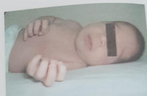
KMT is named by the side on which the head lies. For example, in the right torticollis, the head tilted to the right, the shoulder ear distance decreased, and the face turned to the left. One-third
of thecases have a mass in the sternocleidomastoid muscle.It usually disappears in the first few months. Often fibrous band remains in place . The incidence of epidemiology CMT varies between 0.3-2.0%. This condition, which is rarely seen bilaterally, is more common in men on the right side and at a ratio of 3:2 compared to women. CMT is rare in advanced ages. However, this may be a result of late symptoms of neglected cases or fetal SCM developmental disorder.
Other Characteristic Craniofacial Deformities Accompanying Congenital Muscular Torticollis
- Retraction in the eyebrow and zygoma
- Deviation of jaw and nasal tips
- Inferior orbital dystopia on the affected side
- Inferior and posteriorly located ipsilateral ear
- Unformed craniofacial bone structures
- Shortening of the vertical size of the ipsilateral face
- Sleeping in the supine position, flattening of the cranium on the contralateral side
- Sleeping in the prone position face flattening on the ipsilateral side
- Bend neck position

Mechanism Of Occurrence
1- Abnormal position of the head in the uterus
2- The baby is exposed to any trauma in the uterus
3- Disruption of blood circulation of the NOVEMBERM muscle
4- Rupture of november fibers during childbirth and especially during breech births
5- Primary congenital defect of the cervical vertebrae
The exact cause of congenital muscular torticollis is still a controversial issue. The most accepted theory is compression or vascular phenomenon in the womb. 30-60% of cases of torticollis have a history of difficult childbirth. There are many studies that support the view that torticollis is due to posture abnormalities that can cause compression and a more difficult and traumatic birth in the uterus due to the fact that the uterus is narrower at first birth. November dec november theory, the picture of compression of the neck vessels and the associated compartment syndrome (muscle necrosis due to compression of the muscles between november muscle covers) develops at birth.
Risk Factors
Reverse childbirth
Delayed cesarean section
Twin births
Complicated births
Birth traumas
Muscular torticollis is evaluated in 3 subgroups according to the Macdonald classification:
– The first group, known as fibromatosis colli, is the most common type, which is often accompanied by a hard, mobile tumor located near the point of attachment of the hardened SCM november to the clavicle, painless on palpation, and is the most common type. A november, fusiform-shaped mass with a diameter of 1-3 cm, which can regress in the first year of life, usually noticeable in the first weeks after birth, is observed bilaterally by 2-8%.
– In the second group, which is evaluated as muscular torticollis, there is stiffness in the SCM muscle, but no palpable tumor is detected.
– In the third group called postural torticollis, no mass or stiffness is detected in the SCM muscle.Unlike CMT, normal passive cervical joint range of motion is accompanied by active rotation and intermittent head obstruction with limited lateral flexion.
Evaluation
Before fibrosis occurs in the SCM muscle , all newborns should be examined in detail in order to recognize the disease. Good results are obtained in patients who are diagnosed early and receive conservative treatment as soon as possible, and the need for surgical treatment is very low.The
evaluation starts with anamnesis. Detailed birth history, presence of asymmetry at the beginning and face at birth, palpation of the SCM muscle, physiological movements of the neck and head, and range of motion should be examined. If there is any doubtful movement limitation or obliquity, measurement should be made with an inclinometer and a goniometer. Head tilts with inclinometer or goniometer measurements
- if 1°-15 °, light,
- Medium if 16° – 30°,
- If it is more than 30°, it is defined as heavy.
In addition, the head can be evaluated by measuring on the photograph that fully displays the face and body in the child lying on his/her back. The first line connecting the pupils is drawn on the photo. The second line joining the acromions is drawn and the narrow angle between these two lines is measured by hand.
Bilateral active / passive cervical rotation and lateral flexion degree
Lower extremity abduction asymmetry (to detect hip dysplasia)
Presence of skin folds and rashes on the lateral flexion side of the neck
The thickness of the SCM muscle and the size of the mass within it should also be evaluated.

Although CMT is seen as a problem affecting only the head and neck, functional C scoliosis in the opposite direction of the torticollis side of the body is seen in infants, followed by abduction and external rotation in the leg in the opposite direction of the side causing asymmetry in the pelvis. In addition, the use of extrmeters with torticollis and the decrease in sensory awareness, symmetrical postures in the baby’s body, and weight transfer to one side can be seen. Therefore, it is important to evaluate babies naked.
To exclude ophthalmological and audiological causes, visual field and sound response should be evaluated; radiological and laboratory tests should be performed with multidisciplinary approaches to illuminate the etiology.
Radiological Examination Ultrasound is the most preferred imaging method for radiological evaluation of CMT. In normal SCM muscle ultrasound, it is seen as a hypoechoic area with echogenic lines representing muscle fascicles that pass along the length of the muscle. The presence of SCM tumor affects not only the size of the muscle but also the signal intensity on ultrasound. In CMT, the muscle appears more hyperechogenic. Anteroposterior and lateral cervical radiographs may be requested from patients to rule out cervical vertebral fractures and subluxations.
Magnetic resonance imaging (MRI) can be used to evaluate muscle thickening and fibrosis, posterior fossa tumors. Contrast-enhanced MRI may be requested to rule out cranial tumors.
Computed tomography (CT) or three-dimensional CT can be used to visualize craniofacial and cervical vertebral anomalies. The presence of denervation in the muscle and nerve can be
demonstrated by electromyographic examinations.
Muscle Function Scale (MFS): MFS measures the corrective response of the baby by visual evaluation. The baby is held in the vertical position, then slowly shortened in front of the mirror and placed in the horizontal position towards the sternocleidomastoid muscle side.

The head position is observed and scored over 0-5 points, taking into account the position of the head relative to the vertical line . Infants who startprognosistreatment early will recover in a shorter time. The positional torticollis heals before 3 months. Recovery is faster and shorter in children whose treatment is started before 1 month. It is very important to educate the family so that asymmetry does not develop. CMT is a musculoskeletal condition in which complete recovery can be achieved. However, posture disorders of CMT may be permanent when physiotherapy is started late and not applied effectively.
More than 90% of the cases respond very well to conservative treatment and recover fully. The baby should be evaluated surgically if the mass does not shrink and asymmetry persistently continues despite regular 6-8 months of exercise treatment. The most important factor that determines surgery is that there is a mass in the muscle and the mass is large. Inneglected and/or delayed cases, diagnosis and treatment may progress with increases in the number and size of fibrotic involvement in the SCM muscle.
Serious limitations in the cervical joint range of motion, craniofacial and spinal deformities and neurological losses, which are the basis of this condition, require longer and complex treatment processes and may cause permanent damage that cannot be treated.
In untreated and/or unresponsive cases, deformities such as plagiosephaly, zygoma and ear extraction may develop. Then, the negative effects of the head tilt on the shoulder, rib cage, abdominal muscles, asymmetry in the use of the upper extremity, subnormal cognitive function, posture and balance (sensory-motor coordination) control can be detected.
It has been reported that these changes may be permanent despite corrective surgery in cases after the age of 5.
Physiotherapy in KMT
Cases Where Physiotherapy Is Contraindicated:
Lateral flexion and rotation of less than 5 degrees
Sudden onset torticollis
Tumor of the posterior fossa
Hemiplegia
The presence of fractures and dislocations of the vertebrae
Abnormal vertebral structure (Klippel-Feil Syndrome, hemivertebra)
Abnormal placement of cervical vertebrae
All these are situations where passive cervical movements are prohibited.
Physiotherapy approaches
Family education
Positioning
Stretching exercises
Active exercises and strengthening
Massage
Supporting development
Babies should be followed weekly until 6-8 months. Babies who are in the December of 9-12 months who are admitted late should be seen and monitored once a week again.
Family Education
Family Education Treatment is started with family education.The family is given the necessary information about torticollis; controls and follow-ups, stages of treatment and different treatment options are explained.
Positioning
–The baby should be positioned on the face, back and side during the whole day.
(NOTE: The face should be placed on the sheep and/or sideways for at least 30 minutes 3 times in total during the day, and the time spent in the supine position and car seat should be reduced as much as possible.)
– Shortened SKM is recommended to lie on the muscle side with a thick and hard pillow and on the other side without a pillow.
– 5 times a day, 2-5 minutes should be placed face down on the shortened SKM side. (Toys should be placed on the side where the SKM muscle is affected or games should be played/communicated to adopt the position when lying face down)
– On-back symmetrical lying should be provided in the assembly prepared with a hard and tight peak.
-The rigid external supports used for symmetrical reclining in the car seat and main lap should include not only the head but also the body and pelvis.
– In the baby’s room, stimuli and light should be given from the side where he/she cannot turn his/her neck, toys should be shown from the same


Transportation
When carrying the baby, the torticollis side is supported under the neck with one hand, and the forearm is held at ear level. The other arm is passed between the legs, and the baby’s dec is supported.
The baby is moved with his face outward, while the neck is brought into rotation to the side where he cannot turn. With this grip, it is recommended to carry the baby 5-6 times a day for 5-10 minutes.
The baby should be breastfed with the side of the CMT resting on the mother’s arm.

Passive Stretching
In order to extend the shortened side SKM, lateral flexion to the opposite side is expected for 10 seconds with the face facing the opposite side.
Passive rotation to the shortened side is performed and 10 seconds are expected.
These two exercises can be performed 3-5 times a day for 10-15 repetitions according to the baby’s needs.
The stretching exercise should never be painful, it should be abandoned as soon as there is resistance from the baby. It is noted that low-intensity, continuous, painless stretching will not cause microtraumas.


Active Exercises and Strengthening
Strengthening with the use ofneck correction reaction:
The baby is held from the body and placed in an upright position. Then, towards the KMT side, the body begins to be tilted slowly. The body is taken up to the point where the baby can hold his head upright and then brought back after being held a little at this point. This activity can also be done on the exercise ball if the baby’s developmental step is appropriate. It is recommended to perform active rotation exercises on the side with CMT by drawing the attention of the baby with audible toys duringsupine and prone lying. Exercises should be done 8-10 times every hour when the baby is awake and calm and in a way that does not tire the baby.

Massage
If there is a mass in theshorter side SCM muscle, it should be compressed between the almond oil/baby oil and the thumb and index finger and massage should be performed at
regular intervalsfor 1-2 minutes.
Supporting Development The use of asymmetric movement patterns by babies with torticollis can cause motor development delay and unilateral and different rotational, sitting and creeping movements.For this reason, motor development should be supported by analyzing the movements of the baby with torticollis in a good way. Neuromuscular facilitation techniques can be used to assist in the development ofnormal head position, age-appropriate gross motor function. With these techniques, it is supported that the child tries to hold his/her head in the midline with his/her body on his/her elbows while in the prone position by raising it as much as he/she can. Exercises can be applied to try to stand on four extremities, such as turning on their backs, sitting up, and head in the midline. In order to prevent the development of functional asymmetry and to improve the rotation, capture and sitting suitable for the age, the child should be given appropriate exercises for the
developmental period.

Kinesiological Taping :Kinesiological taping in torticollis is used to fascinate excessively elongated muscles, to correct the baby’s neck and to keep it at the midline.The band facilitates sensory stimulation to the SCM muscle. Supports upright and midline posture. Aftertaping,
the position of the head should be re-evaluated. Tight gluing of the tape causes fatigue and the tape cannot be tolerated.
Banding is initially applied 2-3 times a week with 2 days of banding and 1 day intervals.The application of kinesiological taping to the child with right torticollis is as follows:
After the left lateral flexion and right rotation position are given to the head, taping is performed from the origin of both heads of the left SCM muscle to the insertion.
Orthosis
Orthotic treatment is required in infants with severe or moderate deformational plagiocephaly and / or in infants who do not benefit from classical treatment, in infants with persistent concomitant anterior craniofacial deformities. Orthosis therapy stimulates the reshaping of the head. It is prepared individually. In the cranial orthosis (helmet orthosis), a space is left on the flattening part of the cranium to allow it to take shape, pressure is applied to the areas necessary to ensure symmetry. Every 1-3 weeks, a modification of the orthosis is required. Initially, it is worn for 15-22 hours a day, after 3-4 months it can be worn only at night. This orthosis can be used starting from the age of 4-8 months and ending at the age of 18 months.
Two months after the rehabilitation program, cervical orthosis can be performed in children with no improvement in the lateral rotation of the neck and head tilt, the head tilt is above 16º during the initial examination, while they are awake. If the head tilt is above 16º, soft cervical orthosis is preferred. If the head tilt is above 30°, an orthosis made of thermoplastic material covering the affected side of the face and holding the head in a neutral position can be used. It can be seen that there is no clear indication in the literature on the use of orthotics and no definite conclusion has been reached about the results of its use.

Examples of Physiotherapy Applications in KMT
The exercise ball is tilted to the side of the torticollis, correcting the head on the opposite side.
In order to increase awareness and use of the extremities on the side of the torticollis, weight transfer, a barbed ball and toys of different textures are used.
In positions where the legs come to bilateral flexion, good and symmetrical weight transfer to the neck can be studied.
If the baby is evaluated in terms of movement development, there is rotation only from one side, weight transfer and rotation studies are also added to the program on the other side.
Exercises that work lateral flexion and rotation to the opposite side on the ball can be given as a home program.

 Physiotherapy after surgery After surgery, an orthosis is used that covers the head, neck and shoulders for 3 weeks and holds the shortened side in the opposite lateral flexion and rotation. After the period of continuous use of the orthosis, passive and active neck lateral flexion and rotation exercises are performed. PNF patterns can also be used if there is an active participation of children.
Leave a Reply